File:In-Vivo-Open-Bore-MRI-Reveals-Region--and-Sub-Arc-Specific-Lengthening-of-the-Unloaded-Human-pone.0048714.s001.ogv
Jump to navigation
Jump to search
Size of this JPG preview of this OGG file: 800 × 450 pixels. Other resolutions: 320 × 180 pixels | 640 × 360 pixels | 960 × 540 pixels.
Original file (Ogg Theora video file, length 15 s, 960 × 540 pixels, 1.13 Mbps, file size: 1.97 MB)
Captions
Captions
Add a one-line explanation of what this file represents
Summary
[edit]| Description |
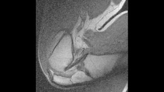
English: Video made from static 2D MR images that were stitched together. Images are para-sagittal with the anterior of the knee to the left and the posterior to the right. The video provides good visualization of many soft tissues, including the PCL, the patellar tendon, Hoffa’s fat pad and the synovium (see Figure 3). |
||
| Date | |||
| Source | Video S1 from King A, Deng Q, Tyson R, Sharp J, Matwiy J, Tomanek B, Dunn J (2012). "In Vivo Open-Bore MRI Reveals Region- and Sub-Arc-Specific Lengthening of the Unloaded Human Posterior Cruciate Ligament". PLOS ONE. DOI:10.1371/journal.pone.0048714. PMID 23144939. PMC: 3492418. | ||
| Author | King A, Deng Q, Tyson R, Sharp J, Matwiy J, Tomanek B, Dunn J | ||
| Permission (Reusing this file) |
|
||
| Provenance |
|
File history
Click on a date/time to view the file as it appeared at that time.
| Date/Time | Thumbnail | Dimensions | User | Comment | |
|---|---|---|---|---|---|
| current | 11:54, 13 November 2012 | 15 s, 960 × 540 (1.97 MB) | Open Access Media Importer Bot (talk | contribs) | Automatically uploaded media file from Open Access source. Please report problems or suggestions here. |
You cannot overwrite this file.
File usage on Commons
There are no pages that use this file.
Transcode status
Update transcode statusFile usage on other wikis
The following other wikis use this file: