Category:Human bones on black background
Jump to navigation
Jump to search
Français : Os humains sur fond noir
Media in category "Human bones on black background"
The following 147 files are in this category, out of 147 total.
-
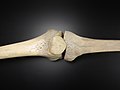 Articulatio genus - frontal.jpg 4,608 × 3,456; 4.26 MB
-
 Articulatio genus - lateral.jpg 4,608 × 3,456; 4.06 MB
-
 Clavicula - dex.jpg 4,608 × 3,456; 5 MB
-
 Clavicula - sin, dex.jpg 4,608 × 3,456; 4.89 MB
-
 Costa - articular facet.jpg 4,608 × 3,456; 4.12 MB
-
 Costae - 1, 2, 3.jpg 4,608 × 3,456; 4.31 MB
-
 Costae - articular facets.jpg 4,608 × 3,456; 4.58 MB
-
 Costae - collection.jpg 4,608 × 3,456; 4.43 MB
-
 Costae - dex.jpg 4,608 × 3,456; 4.59 MB
-
 Costae - sin, dex.jpg 4,608 × 3,456; 4.32 MB
-
 Crane1.png 2,800 × 2,662; 5.47 MB
-
 Crane3chin.png 1,404 × 1,924; 2.71 MB
-
 Crane4 Foramen magnum.png 2,431 × 2,761; 6.83 MB
-
 Crane4.png 2,431 × 2,761; 6.96 MB
-
.jpg/120px-Cranium_-_basis_cranii_interna%2C_maxilla_(lateral).jpg) Cranium - basis cranii interna, maxilla (lateral).jpg 4,608 × 3,456; 4.1 MB
Cranium - basis cranii interna, maxilla (lateral).jpg 4,608 × 3,456; 4.1 MB
-
 Cranium - inferior view with atlas.jpg 3,816 × 2,816; 3.59 MB
-
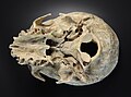
 Cranium - inferior view.jpg 4,380 × 3,396; 5.43 MB
-
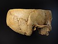
 Cranium - lateral view 2.jpg 4,608 × 3,456; 3.52 MB
-
 Cranium - lateral view.jpg 4,608 × 3,456; 4.79 MB
-
.jpg/90px-Cranium_-_mandibula_(anterior).jpg) Cranium - mandibula (anterior).jpg 3,456 × 4,608; 3.46 MB
Cranium - mandibula (anterior).jpg 3,456 × 4,608; 3.46 MB
-
.jpg/120px-Cranium_-_mandibula_(dental_bridge%2C_dentistry).jpg) Cranium - mandibula (dental bridge, dentistry).jpg 4,608 × 3,456; 4.49 MB
Cranium - mandibula (dental bridge, dentistry).jpg 4,608 × 3,456; 4.49 MB
-
.jpg/120px-Cranium_-_mandibula_(lateral_view).jpg) Cranium - mandibula (lateral view).jpg 4,608 × 3,456; 3.62 MB
Cranium - mandibula (lateral view).jpg 4,608 × 3,456; 3.62 MB
-
.jpg/90px-Cranium_-_mandibula_(toothless).jpg) Cranium - mandibula (toothless).jpg 3,456 × 4,608; 4.21 MB
Cranium - mandibula (toothless).jpg 3,456 × 4,608; 4.21 MB
-
 Cranium - neurocranium posterior view 2.jpg 3,456 × 4,608; 4.38 MB
-
 Cranium - neurocranium posterior view 3.jpg 3,456 × 4,608; 4.38 MB
-
 Cranium - neurocranium posterior view.jpg 3,456 × 4,608; 4.49 MB
-
.jpg/120px-Cranium_-_neurocranium_superior_view_(sutures).jpg) Cranium - neurocranium superior view (sutures).jpg 4,608 × 3,456; 4.55 MB
Cranium - neurocranium superior view (sutures).jpg 4,608 × 3,456; 4.55 MB
-
 Cranium - neurocranium superior view 2.jpg 4,608 × 3,456; 4.66 MB
-
 Cranium - neurocranium superior view.jpg 3,456 × 4,608; 3.34 MB
-
.jpg/120px-Cranium_-_os_occipitale_(external).jpg) Cranium - os occipitale (external).jpg 4,608 × 3,456; 4.69 MB
Cranium - os occipitale (external).jpg 4,608 × 3,456; 4.69 MB
-
.jpg/120px-Cranium_-_os_occipitale_(internal).jpg) Cranium - os occipitale (internal).jpg 4,608 × 3,456; 4.05 MB
Cranium - os occipitale (internal).jpg 4,608 × 3,456; 4.05 MB
-
.jpg/120px-Cranium_-_os_parietale_(inner_side).jpg) Cranium - os parietale (inner side).jpg 4,608 × 3,456; 5.05 MB
Cranium - os parietale (inner side).jpg 4,608 × 3,456; 5.05 MB
-
 Cranium - pterion.jpg 3,648 × 2,936; 3.83 MB
-
_2.jpg/90px-Cranium_-_splanchnocranium_(anterior_view)_2.jpg) Cranium - splanchnocranium (anterior view) 2.jpg 3,456 × 4,608; 4.77 MB
Cranium - splanchnocranium (anterior view) 2.jpg 3,456 × 4,608; 4.77 MB
-
.jpg/90px-Cranium_-_splanchnocranium_(anterior_view).jpg) Cranium - splanchnocranium (anterior view).jpg 3,456 × 4,608; 4.33 MB
Cranium - splanchnocranium (anterior view).jpg 3,456 × 4,608; 4.33 MB
-
.jpg/90px-Cranium_-_sutura_frontalis_metopica_(anterior).jpg) Cranium - sutura frontalis metopica (anterior).jpg 3,456 × 4,608; 3.17 MB
Cranium - sutura frontalis metopica (anterior).jpg 3,456 × 4,608; 3.17 MB
-
.jpg/120px-Cranium_-_sutura_frontalis_metopica_(posterior).jpg) Cranium - sutura frontalis metopica (posterior).jpg 4,608 × 3,456; 5.06 MB
Cranium - sutura frontalis metopica (posterior).jpg 4,608 × 3,456; 5.06 MB
-
 CTSkullImage - cropped.jpg 768 × 600; 62 KB
-
 CTSkullImage.png 580 × 580; 336 KB
-
 Detail of human bone tissue.jpg 3,312 × 2,496; 3.56 MB
-
_-_bone_structure_detail_(vertical_cut)_2.jpg/120px-Femur_(caput_femoris)_-_bone_structure_detail_(vertical_cut)_2.jpg) Femur (caput femoris) - bone structure detail (vertical cut) 2.jpg 4,608 × 3,456; 4.45 MB
Femur (caput femoris) - bone structure detail (vertical cut) 2.jpg 4,608 × 3,456; 4.45 MB
-
_-_bone_structure_detail_(vertical_cut)_3.jpg/120px-Femur_(caput_femoris)_-_bone_structure_detail_(vertical_cut)_3.jpg) Femur (caput femoris) - bone structure detail (vertical cut) 3.jpg 4,168 × 2,348; 4.08 MB
Femur (caput femoris) - bone structure detail (vertical cut) 3.jpg 4,168 × 2,348; 4.08 MB
-
_-_bone_structure_detail_(vertical_cut).jpg/90px-Femur_(caput_femoris)_-_bone_structure_detail_(vertical_cut).jpg) Femur (caput femoris) - bone structure detail (vertical cut).jpg 3,456 × 4,608; 3.7 MB
Femur (caput femoris) - bone structure detail (vertical cut).jpg 3,456 × 4,608; 3.7 MB
-
.jpg/120px-Femur_-_anterior_view_(sin%2C_dex).jpg) Femur - anterior view (sin, dex).jpg 4,232 × 2,384; 3.14 MB
Femur - anterior view (sin, dex).jpg 4,232 × 2,384; 3.14 MB
-
 Femur - anterior, posterior.jpg 4,240 × 3,272; 4.75 MB
-
.jpg/103px-Femur_-_caput_femoris_(anterior).jpg) Femur - caput femoris (anterior).jpg 2,604 × 3,024; 2.88 MB
Femur - caput femoris (anterior).jpg 2,604 × 3,024; 2.88 MB
-
.jpg/105px-Femur_-_caput_femoris_(posterior).jpg) Femur - caput femoris (posterior).jpg 2,664 × 3,056; 3.1 MB
Femur - caput femoris (posterior).jpg 2,664 × 3,056; 3.1 MB
-
 Femur - detail of diaphysis cross section.jpg 3,042 × 2,652; 2.64 MB
-
.jpg/120px-Femur_-_epicondylus_lateralis_et_medialis_(posterior_view).jpg) Femur - epicondylus lateralis et medialis (posterior view).jpg 4,608 × 3,456; 4.48 MB
Femur - epicondylus lateralis et medialis (posterior view).jpg 4,608 × 3,456; 4.48 MB
-
.jpg/120px-Femur_-_posterior_view_(sin%2C_dex).jpg) Femur - posterior view (sin, dex).jpg 4,372 × 2,380; 3.75 MB
Femur - posterior view (sin, dex).jpg 4,372 × 2,380; 3.75 MB
-
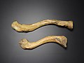
.jpg/120px-Fibula_(anterior%2C_posterior).jpg) Fibula (anterior, posterior).jpg 3,780 × 1,638; 2.13 MB
Fibula (anterior, posterior).jpg 3,780 × 1,638; 2.13 MB
-
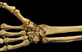
 Handskelett 55j weiblich.png 562 × 1,084; 275 KB
-
 Handskelett 66j maennlich.png 638 × 1,088; 343 KB
-
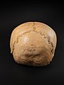
 Human Skull.jpg 1,844 × 2,474; 546 KB
-
 Humerus - anterior 2.jpg 4,488 × 2,288; 3.18 MB
-
 Humerus - anterior.jpg 4,448 × 1,944; 2.68 MB
-
 Humerus - articulatio humeri.jpg 4,608 × 3,456; 5.24 MB
-
 Humerus - posterior 2.jpg 4,608 × 3,456; 4.34 MB
-
 Humerus - posterior.jpg 4,272 × 2,496; 3.19 MB
-
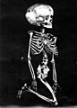 Hydrocephalic skeleton.jpg 844 × 1,180; 601 KB
-
 Myrtis skull.jpg 768 × 1,024; 228 KB
-
.jpg/120px-Nyamata_Memorial_Site_13_(cropped).jpg) Nyamata Memorial Site 13 (cropped).jpg 2,466 × 1,690; 1.4 MB
Nyamata Memorial Site 13 (cropped).jpg 2,466 × 1,690; 1.4 MB
-
 Nyamata Memorial Site 13-Version 2.png 3,008 × 2,000; 3.67 MB
-
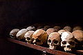
 Nyamata Memorial Site 13.jpg 3,008 × 2,000; 1.71 MB
-
 Nyamata Memorial Site 13b.jpg 3,008 × 2,000; 1.03 MB
-
_-_calcaneus_(facies_articularis_talaris).jpg/120px-Ossa_pedis_(sin)_-_calcaneus_(facies_articularis_talaris).jpg) Ossa pedis (sin) - calcaneus (facies articularis talaris).jpg 4,608 × 3,456; 4.64 MB
Ossa pedis (sin) - calcaneus (facies articularis talaris).jpg 4,608 × 3,456; 4.64 MB
-
_-_calcaneus_(medial_view_detail).jpg/120px-Ossa_pedis_(sin)_-_calcaneus_(medial_view_detail).jpg) Ossa pedis (sin) - calcaneus (medial view detail).jpg 4,608 × 3,456; 3.99 MB
Ossa pedis (sin) - calcaneus (medial view detail).jpg 4,608 × 3,456; 3.99 MB
-
_-_calcaneus_(medial_view).jpg/120px-Ossa_pedis_(sin)_-_calcaneus_(medial_view).jpg) Ossa pedis (sin) - calcaneus (medial view).jpg 4,608 × 3,456; 4.53 MB
Ossa pedis (sin) - calcaneus (medial view).jpg 4,608 × 3,456; 4.53 MB
-
_-_lateral_view.jpg/120px-Ossa_pedis_(sin)_-_lateral_view.jpg) Ossa pedis (sin) - lateral view.jpg 4,608 × 3,456; 4.61 MB
Ossa pedis (sin) - lateral view.jpg 4,608 × 3,456; 4.61 MB
-
_-_medialis_et_superior.jpg/120px-Ossa_pedis_(sin)_-_medialis_et_superior.jpg) Ossa pedis (sin) - medialis et superior.jpg 4,608 × 4,424; 3.33 MB
Ossa pedis (sin) - medialis et superior.jpg 4,608 × 4,424; 3.33 MB
-
_-_ossa_metatarsi_(medial_view).jpg/120px-Ossa_pedis_(sin)_-_ossa_metatarsi_(medial_view).jpg) Ossa pedis (sin) - ossa metatarsi (medial view).jpg 4,608 × 3,456; 4.01 MB
Ossa pedis (sin) - ossa metatarsi (medial view).jpg 4,608 × 3,456; 4.01 MB
-
_-_ossa_metatarsi_(superior_view).jpg/120px-Ossa_pedis_(sin)_-_ossa_metatarsi_(superior_view).jpg) Ossa pedis (sin) - ossa metatarsi (superior view).jpg 4,608 × 3,456; 4.61 MB
Ossa pedis (sin) - ossa metatarsi (superior view).jpg 4,608 × 3,456; 4.61 MB
-
_-_superior_view.jpg/120px-Ossa_pedis_(sin)_-_superior_view.jpg) Ossa pedis (sin) - superior view.jpg 4,608 × 3,456; 3.11 MB
Ossa pedis (sin) - superior view.jpg 4,608 × 3,456; 3.11 MB
-
.jpg/120px-Ossa_pedis_-_talus_(medialis%2C_lateralis).jpg) Ossa pedis - talus (medialis, lateralis).jpg 6,976 × 3,456; 8.63 MB
Ossa pedis - talus (medialis, lateralis).jpg 6,976 × 3,456; 8.63 MB
-
.jpg/120px-Ossa_pedis_-_talus_(superior_view).jpg) Ossa pedis - talus (superior view).jpg 3,176 × 2,472; 3.02 MB
Ossa pedis - talus (superior view).jpg 3,176 × 2,472; 3.02 MB
-
 Ossuary in Sedlec.JPG 2,002 × 1,442; 1.99 MB
-
 Patella - detail of bone tissue.jpg 3,760 × 2,768; 2.05 MB
-
 Patella - facies anterior et posterior patellae.jpg 4,164 × 3,456; 5.94 MB
-
 Patella - facies anterior patellae detail.jpg 4,608 × 3,456; 4.63 MB
-
 Patella - facies anterior patellae.jpg 4,608 × 3,456; 4.46 MB
-
 Patella - facies posterior patellae detail.jpg 4,608 × 3,456; 4.3 MB
-
 Patella - facies posterior patellae.jpg 4,608 × 3,456; 4.28 MB
-
.jpg/120px-Pelvis_-_acetabulum_(os_coxae).jpg) Pelvis - acetabulum (os coxae).jpg 4,608 × 3,456; 5 MB
Pelvis - acetabulum (os coxae).jpg 4,608 × 3,456; 5 MB
-
.jpg/120px-Pelvis_-_os_coxae_(dex%2C_sin).jpg) Pelvis - os coxae (dex, sin).jpg 4,608 × 3,456; 5.03 MB
Pelvis - os coxae (dex, sin).jpg 4,608 × 3,456; 5.03 MB
-
_2.jpg/120px-Pelvis_-_os_coxae_(lateral_view)_2.jpg) Pelvis - os coxae (lateral view) 2.jpg 4,608 × 3,456; 4.4 MB
Pelvis - os coxae (lateral view) 2.jpg 4,608 × 3,456; 4.4 MB
-
.jpg/90px-Pelvis_-_os_coxae_(lateral_view).jpg) Pelvis - os coxae (lateral view).jpg 3,456 × 4,608; 4.71 MB
Pelvis - os coxae (lateral view).jpg 3,456 × 4,608; 4.71 MB
-
.jpg/120px-Pelvis_-_os_coxae_-_dex%2C_sin_(lateral).jpg) Pelvis - os coxae - dex, sin (lateral).jpg 4,608 × 3,456; 4.92 MB
Pelvis - os coxae - dex, sin (lateral).jpg 4,608 × 3,456; 4.92 MB
-
.jpg/120px-Pelvis_-_os_coxae%2C_os_sacrum_(caudal_view).jpg) Pelvis - os coxae, os sacrum (caudal view).jpg 4,608 × 3,456; 4.54 MB
Pelvis - os coxae, os sacrum (caudal view).jpg 4,608 × 3,456; 4.54 MB
-
.jpg/120px-Pelvis_-_os_coxae%2C_os_sacrum_(frontal_view).jpg) Pelvis - os coxae, os sacrum (frontal view).jpg 4,608 × 3,456; 4.47 MB
Pelvis - os coxae, os sacrum (frontal view).jpg 4,608 × 3,456; 4.47 MB
-
.jpg/120px-Pelvis_-_os_coxae%2C_os_sacrum_(lateral_view).jpg) Pelvis - os coxae, os sacrum (lateral view).jpg 3,440 × 2,604; 2.66 MB
Pelvis - os coxae, os sacrum (lateral view).jpg 3,440 × 2,604; 2.66 MB
-
.jpg/120px-Pelvis_-_os_coxae%2C_os_sacrum_(posterior_view).jpg) Pelvis - os coxae, os sacrum (posterior view).jpg 4,140 × 2,712; 3.48 MB
Pelvis - os coxae, os sacrum (posterior view).jpg 4,140 × 2,712; 3.48 MB
-
 Pelvis - os coxae, os sacrum.jpg 4,608 × 3,456; 4.3 MB
-
.jpg/120px-Pelvis_-_os_ilium_(posterior).jpg) Pelvis - os ilium (posterior).jpg 4,608 × 3,456; 5.07 MB
Pelvis - os ilium (posterior).jpg 4,608 × 3,456; 5.07 MB
-
 Pelvis - os illium - detail of bone tissue.jpg 4,288 × 2,704; 4.01 MB
-
.jpg/120px-Pelvis_-_os_ischii%2C_acetabulum_(lateral).jpg) Pelvis - os ischii, acetabulum (lateral).jpg 4,608 × 3,456; 5.2 MB
Pelvis - os ischii, acetabulum (lateral).jpg 4,608 × 3,456; 5.2 MB
-
.jpg/97px-Pelvis_-_os_sacrum_(anterior).jpg) Pelvis - os sacrum (anterior).jpg 2,460 × 3,048; 3.43 MB
Pelvis - os sacrum (anterior).jpg 2,460 × 3,048; 3.43 MB
-
.jpg/120px-Pelvis_-_os_sacrum_(caudal_view).jpg) Pelvis - os sacrum (caudal view).jpg 3,844 × 2,492; 4.26 MB
Pelvis - os sacrum (caudal view).jpg 3,844 × 2,492; 4.26 MB
-
.jpg/120px-Pelvis_-_os_sacrum_(lateral_view).jpg) Pelvis - os sacrum (lateral view).jpg 3,692 × 2,320; 3.71 MB
Pelvis - os sacrum (lateral view).jpg 3,692 × 2,320; 3.71 MB
-
_2.jpg/120px-Pelvis_-_os_sacrum_(posterior)_2.jpg) Pelvis - os sacrum (posterior) 2.jpg 3,720 × 2,496; 3.71 MB
Pelvis - os sacrum (posterior) 2.jpg 3,720 × 2,496; 3.71 MB
-
.jpg/103px-Pelvis_-_os_sacrum_(posterior).jpg) Pelvis - os sacrum (posterior).jpg 2,748 × 3,192; 3.18 MB
Pelvis - os sacrum (posterior).jpg 2,748 × 3,192; 3.18 MB
-
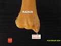
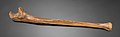
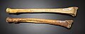 Radius - anterior.jpg 4,108 × 1,676; 2.7 MB
-
.jpg/120px-Radius_-_detail_of_bone_tissue_(distal_end).jpg) Radius - detail of bone tissue (distal end).jpg 3,320 × 2,840; 3.73 MB
Radius - detail of bone tissue (distal end).jpg 3,320 × 2,840; 3.73 MB
-
 Radius - posterior 2.jpg 4,608 × 3,456; 4.65 MB
-
 Radius - posterior.jpg 4,320 × 1,752; 2.7 MB
-
 Radius3.jpg 960 × 720; 42 KB
-
 Radius4.jpg 960 × 720; 49 KB
-
 RightHumanAnteriorDistalRadiusUlnaCarpals.jpg 2,661 × 1,681; 687 KB
-
 Sapiens neanderthal comparison en blackbackground.png 1,025 × 533; 354 KB
-
.jpg/90px-Scapula_-_anterior_(damaged_bone).jpg) Scapula - anterior (damaged bone).jpg 3,456 × 4,608; 4.95 MB
Scapula - anterior (damaged bone).jpg 3,456 × 4,608; 4.95 MB
-
 Scapula - anterior view.jpg 3,456 × 4,608; 4.59 MB
-
 Scapula - coracoid, acromion.jpg 3,456 × 4,608; 4.45 MB
-
 Scapula - posterior view 2.jpg 4,608 × 3,456; 4.43 MB
-
 Scapula - posterior view.jpg 3,456 × 4,608; 4.6 MB
-
.jpg/90px-Scapula%2C_clavicula_-_posterior_(model).jpg) Scapula, clavicula - posterior (model).jpg 3,456 × 4,608; 4.17 MB
Scapula, clavicula - posterior (model).jpg 3,456 × 4,608; 4.17 MB
-
 Small-Bodied Humans from Palau, Micronesia - Bones.png 303 × 270; 53 KB
-
 Tibia - condylus lateralis et medialis.jpg 4,608 × 3,456; 4.8 MB
-
_2.jpg/120px-Tibia_-_detail_of_bone_tissue_(proximal_end)_2.jpg) Tibia - detail of bone tissue (proximal end) 2.jpg 3,976 × 3,008; 4.44 MB
Tibia - detail of bone tissue (proximal end) 2.jpg 3,976 × 3,008; 4.44 MB
-
.jpg/120px-Tibia_-_detail_of_bone_tissue_(proximal_end).jpg) Tibia - detail of bone tissue (proximal end).jpg 4,608 × 3,456; 5.3 MB
Tibia - detail of bone tissue (proximal end).jpg 4,608 × 3,456; 5.3 MB
-
 Tibia - detail of malleolus medialis bone tissue.jpg 2,976 × 2,784; 2.98 MB
-
 Tibia - dex, sin.jpg 4,608 × 3,456; 4.62 MB
-
.jpg/120px-Tibia_-_facies_articularis_superior_(dex%2C_sin).jpg) Tibia - facies articularis superior (dex, sin).jpg 4,320 × 2,408; 3.4 MB
Tibia - facies articularis superior (dex, sin).jpg 4,320 × 2,408; 3.4 MB
-
 Tibia - facies articularis superior 2.jpg 2,932 × 2,108; 2.43 MB
-
 Tibia - facies articularis superior.jpg 3,408 × 2,676; 3.42 MB
-
.jpg/120px-Tibia_-_malleolus_medialis_(dex%2C_sin).jpg) Tibia - malleolus medialis (dex, sin).jpg 3,832 × 2,472; 3.2 MB
Tibia - malleolus medialis (dex, sin).jpg 3,832 × 2,472; 3.2 MB
-
 Tibia - malleolus medialis.jpg 4,608 × 3,456; 4.38 MB
-
.jpg/120px-Tibia%2C_fibula_(anterior).jpg) Tibia, fibula (anterior).jpg 3,860 × 1,696; 2.28 MB
Tibia, fibula (anterior).jpg 3,860 × 1,696; 2.28 MB
-
 Tibia.jpg 4,384 × 2,648; 3.51 MB
-
 Télviec Crane Profil Droit.jpg 2,964 × 2,852; 4.61 MB
-
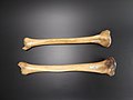
 Ulna - lateral 2.jpg 4,480 × 1,392; 1.9 MB
-
 Ulna - lateral, anterior 2.jpg 4,608 × 3,456; 4.48 MB
-
 Ulna - lateral, anterior.jpg 4,224 × 2,400; 3.97 MB
-
 Ulna - lateral.jpg 4,608 × 3,456; 4.56 MB
-
.jpg/120px-Ulna_-_olecranon_(anterior%2C_lateral).jpg) Ulna - olecranon (anterior, lateral).jpg 3,536 × 3,184; 4.92 MB
Ulna - olecranon (anterior, lateral).jpg 3,536 × 3,184; 4.92 MB
-
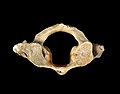 Vertebra - atlas.jpg 3,080 × 2,404; 587 KB
-
.jpg/90px-Vertebra_-_axis_(posterior).jpg) Vertebra - axis (posterior).jpg 3,456 × 4,608; 3.64 MB
Vertebra - axis (posterior).jpg 3,456 × 4,608; 3.64 MB
-
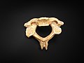 Vertebra - axis.jpg 4,608 × 3,456; 4.22 MB
-
.jpg/120px-Vertebra_-_cervicales_(anterior).jpg) Vertebra - cervicales (anterior).jpg 4,608 × 3,456; 3.75 MB
Vertebra - cervicales (anterior).jpg 4,608 × 3,456; 3.75 MB
-
.jpg/120px-Vertebra_-_cervicales_(from_below).jpg) Vertebra - cervicales (from below).jpg 4,608 × 3,456; 3.72 MB
Vertebra - cervicales (from below).jpg 4,608 × 3,456; 3.72 MB
-
.jpg/120px-Vertebra_-_cervicales_(posterior).jpg) Vertebra - cervicales (posterior).jpg 4,608 × 3,456; 3.8 MB
Vertebra - cervicales (posterior).jpg 4,608 × 3,456; 3.8 MB
-
 Vertebra - cervicales.jpg 3,156 × 2,976; 2.77 MB
-
.jpg/120px-Vertebra_-_lumbales_(superior_view).jpg) Vertebra - lumbales (superior view).jpg 4,608 × 3,456; 3.59 MB
Vertebra - lumbales (superior view).jpg 4,608 × 3,456; 3.59 MB
-
.jpg/120px-Vertebra_-_lumbales_(superior).jpg) Vertebra - lumbales (superior).jpg 4,608 × 3,456; 3.51 MB
Vertebra - lumbales (superior).jpg 4,608 × 3,456; 3.51 MB
-
.jpg/120px-Vertebra_-_thoracicae_(from_below).jpg) Vertebra - thoracicae (from below).jpg 4,608 × 3,456; 4.58 MB
Vertebra - thoracicae (from below).jpg 4,608 × 3,456; 4.58 MB
-
.jpg/120px-Vertebra_-_thoracicae_(top_view).jpg) Vertebra - thoracicae (top view).jpg 4,608 × 3,456; 4.61 MB
Vertebra - thoracicae (top view).jpg 4,608 × 3,456; 4.61 MB
-
 Vertebrae - lumbales.jpg 4,608 × 3,456; 4.52 MB
-
 VisHumanCT-Anaglyph-1.png 1,599 × 1,107; 1.24 MB
-
 VisHumanCT-Anaglyph-2.png 1,599 × 1,107; 771 KB


.jpg)


.jpg)
.jpg)
.jpg)
.jpg)


.jpg)


.jpg)
.jpg)
.jpg)

_2.jpg)
.jpg)
.jpg)
.jpg)


_-_bone_structure_detail_(vertical_cut)_2.jpg)
_-_bone_structure_detail_(vertical_cut)_3.jpg)
_-_bone_structure_detail_(vertical_cut).jpg)
.jpg)

.jpg)
.jpg)

.jpg)
.jpg)
.jpg)


.jpg)


_-_calcaneus_(facies_articularis_talaris).jpg)
_-_calcaneus_(medial_view_detail).jpg)
_-_calcaneus_(medial_view).jpg)
_-_lateral_view.jpg)
_-_medialis_et_superior.jpg)
_-_ossa_metatarsi_(medial_view).jpg)
_-_ossa_metatarsi_(superior_view).jpg)
_-_superior_view.jpg)
.jpg)
.jpg)


.jpg)
.jpg)
_2.jpg)
.jpg)
.jpg)
.jpg)
.jpg)
.jpg)
.jpg)

.jpg)

.jpg)
.jpg)
.jpg)
.jpg)
_2.jpg)
.jpg)
.jpg)


.jpg)


.jpg)


_2.jpg)
.jpg)


.jpg)


.jpg)

.jpg)


.jpg)

.jpg)

.jpg)
.jpg)
.jpg)

.jpg)
.jpg)
.jpg)
.jpg)


